More information is available on the visiting page

Echocardiogram
An echocardiogram is also known as ultrasound examination or sound wave picture of the heart. It uses the same technology that is used to take pictures of the foetus in pregnant women.
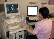
The pictures are taken by a highly trained technician who places a hand-held plastic ultrasound probe against the patient's chest. This is connected to a large computer with a video screen. The probe emits sound waves that pass through the chest to the heart. The heart then reflects those sound waves back to it. The probe transmits those reflected signals to the computer which reconstructs them into a picture of the heart. This picture is displayed on the screen and recorded on videotape or on a digital storage medium.
There are no known harmful side effects from these sound waves. The test does not hurt because normally no needles are used and nothing else is placed inside the patient. The average test takes about 30 minutes.
Sometimes, an ultrasonic contrast agent is injected intravenously to enhance the quality of the sound wave pictures and to give additional information about the amount of blood flow to the heart muscle. Ultrasonic contrast agents are solutions of micro-bubbles. The surface of the bubble is made out of a human protein called albumin. The bubbles contain an inert gas. Side effects are rare, brief and quite mild.
No patient preparation is required for an echocardiogram. The test can be done anywhere, at any time. You are even allowed to eat before this test!
Doctors obtain a wealth of valuable information about the heart from this test at no risk to the patient, and without causing any undue discomfort. Due to its ease, value and safety, the numbers of echocardiogram exams of the heart done in the UK is increasing every year.
An echocardiogram shows:
- The sizes of the 4 chambers of the heart
- The strength of the heart muscle. This is invaluable to doctors in assessing the damage done by heart attacks and in determining how to best treat a patient with heart failure. A lot of the decisions about which medications to give cardiac patients are based on this information
- The presence of fluid around the heart. This condition, known as a pericardial effusion, can progress to a life threatening condition called cardiac tamponade. An echocardiogram is the quickest way to diagnose this condition to avoid delays in treatment
- Problems with the valves of the heart. The heart has 4 valves which must open and close properly in order for the heart to function normally. Abnormal valve function can lead to heart murmurs, difficulty breathing, swelling, and fainting or chest discomfort. Echocardiograms not only detect the presence of these problems but also measure their severity
- Congenital heart disease. Babies born with holes in their hearts or abnormal connections between the heart chambers can be accurately diagnosed with an echocardiogram. It can even be done on the unborn foetus to make a diagnosis so the doctors are ready when the baby is born
- Information about the pressures within the chambers of the heart. Previously, this information could only be obtained by placing catheters into the heart
- Information about why a person may have an erratic heartbeat.
Echocardiogram does not give a picture of the arteries of the heart. Other types of tests are required to diagnose this condition. An echocardiogram, however, can give indirect evidence that a person has blocked arteries by examining the strength of the heart muscle. Ultrasonic contrast agents can also show the amount of blood the heart muscle is receiving.