More information is available on the visiting page

Stress Tests
It is quite common for people who have heart disease to show normal results on heart tests performed under resting, relaxed conditions. However, these results can become abnormal when the heart is made to work harder. This is the basis for stress tests.
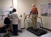
The most common way to stress a person’s heart is to have them perform exercise. Walking on a treadmill is most frequently used. Finally, for patients who are unable to exercise, intravenous medicines can be given to make the heart work as hard as if the person was exercising.
All stress tests are performed with continuous monitoring of the electrocardiogram (ECG). As the heart works harder, certain characteristic abnormalities can develop in the ECG in patients who harbour underlying heart disease.
The exercise, or medication infusion, is performed under the supervision of a health professional.
Sometimes, pictures are taken of the heart in addition to the ECG. These can be echocardiograms or nuclear images. Echo pictures are taken just before the stress (exercise or medicine) begins and again at the peak stress level. This only adds a few minutes to the total duration of the test. The strength of contraction of the heart muscle is compared between the resting and stress echo pictures to determine if an abnormality exists.
Nuclear images are obtained by injecting a mildly radioactive substance that has no known side effects, into a vein in the arm. The bloodstream carries these substances to the heart muscle in proportion to the amount of blood flow to the heart muscle. These substances emit gamma rays which are detected by a camera that constructs a picture of the heart. The injection is given and pictures taken under resting conditions and again following the stress. The pictures show the amount of blood flow to each region of the heart muscle as well as the strength of contraction of the heart muscle.
As with the stress echo, the rest images are compared to the stress images to determine if a heart problem exists. The patient must lie under a camera for between 20 to 45 minutes for each picture to be acquired. Thus, this takes much longer than the other types of stress tests.
The only preparation for a stress test is that you must only eat a light meal, avoid strenuous exercise for 4 hours preceding the test and avoid or minimise smoking for 24 hours beforehand. If it is an exercise test, make sure you wear loose fitting comfortable clothes and good running shoes. High heels don’t work on a treadmill! Your doctor may want you to miss out some of your usual medications on the day of the test. Be sure to check this with your doctor.
Some of the most common reasons for tests being undertaken are:
- To see if a person with no symptoms has silent coronary artery disease, i.e. blockages or hardening of the arteries of the heart. (In many patients, the first signs of heart disease is a heart attack or death without any preceding warning symptoms)
- To determine if a patient’s symptoms (such as chest discomfort or difficulty in breathing) are due to heart disease rather than another problem
- To check that the treatment given a patient for blocked arteries is working properly
- To assess a patient’s exercise tolerance before beginning an exercise or Cardiac Rehabilitation programme
- To determine if exercise causes an abnormal heart rhythm.
Sometimes the results of a Stress Test can indicate there is an issue when the heart is really okay and sometimes they come out as normal when there really is a problem with the heart. Taking echo or nuclear pictures with the stress test makes it more accurate. Whether further testing for blocked arteries beyond a stress test is required depends on each patient's particular circumstances.